ZONES IN THE KIDNEY
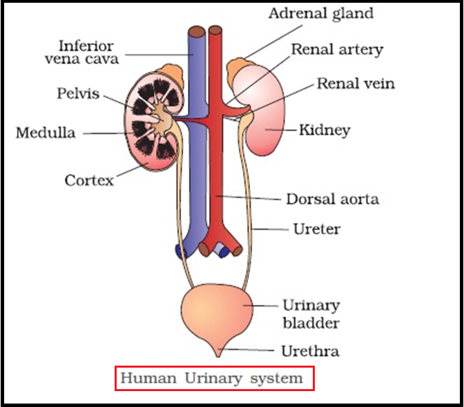
● In humans, the `color{violet}("excretory system")` consists of a `color{brown}("pair of kidneys, one pair of ureters, a urinary bladder")` and a `color{brown}("urethra.")`
● `color{brown}("Kidneys")` are `color{violet}("reddish brown, bean shaped structures situated")` between the levels of last `color{violet}("thoracic and third lumbar vertebra")` close to the `color{violet}("dorsal inner wall")` of the `color{violet}("abdominal cavity.")`
● Each `color{violet}("kidney")` of an adult human measures 10-12 cm in length, 5-7 cm in width, 2-3 cm in thickness with an average weight of 120- 170 g.
● Towards the centre of the inner concave surface of the `color{violet}("kidney")` is a notch called `color{brown}("hilum")` through which ureter, `color{violet}("blood vessels")` and `color{violet}("nerves enter.")`

● Inner to the `color{violet}("hilum")` is a broad funnel shaped space called the `color{brown}("renal pelvis")` with projections called `color{brown}("calyces.")`
● The outer layer of `color{violet}("kidney")` is a `color{brown}("tough capsule.")`
● Inside the `color{violet}("kidney,")` there are two zones, an outer `color{brown}("𝘤𝘰𝘳𝘵𝘦𝘹")` and an inner `color{brown}("𝘮𝘦𝘥𝘶𝘭𝘭𝘢.")`
● The `color{violet}("medulla")` is divided into a few conical masses (`color{brown}("medullary pyramids")`) projecting into the `color{violet}("calyces (sing.: calyx)")`.
● The `color{violet}("cortex extends")` in between the `color{violet}("medullary pyramids")` as `color{violet}("renal columns")` called `color{brown}("Columns of Bertini.")`
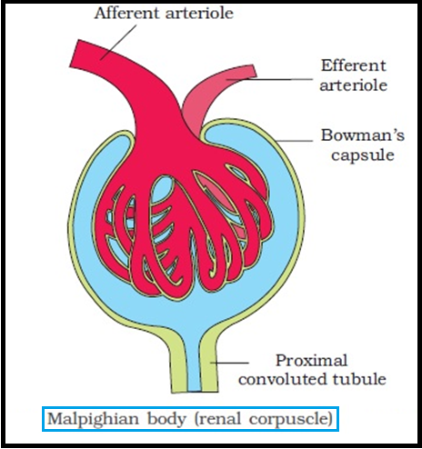
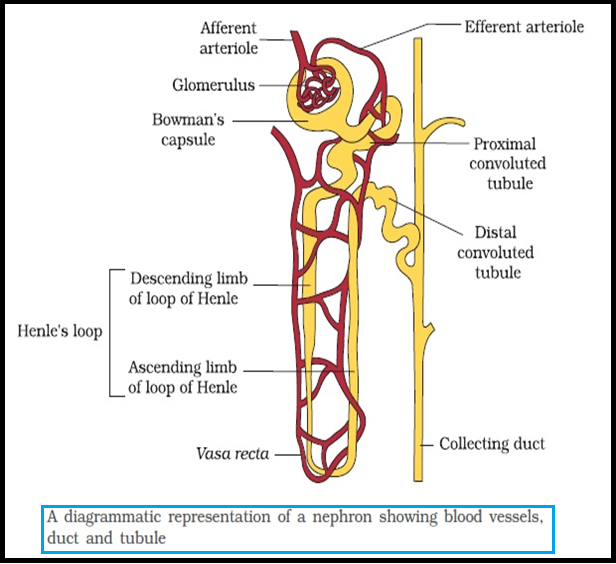
● Each `color{violet}("kidney")` has nearly one million `color{violet}("complex tubular structures")` called `color{brown}("nephrons")` , which are the `color{violet}("functional units.")`
● `color{brown}("Kidneys")` are `color{violet}("reddish brown, bean shaped structures situated")` between the levels of last `color{violet}("thoracic and third lumbar vertebra")` close to the `color{violet}("dorsal inner wall")` of the `color{violet}("abdominal cavity.")`
● Each `color{violet}("kidney")` of an adult human measures 10-12 cm in length, 5-7 cm in width, 2-3 cm in thickness with an average weight of 120- 170 g.
● Towards the centre of the inner concave surface of the `color{violet}("kidney")` is a notch called `color{brown}("hilum")` through which ureter, `color{violet}("blood vessels")` and `color{violet}("nerves enter.")`
● Inner to the `color{violet}("hilum")` is a broad funnel shaped space called the `color{brown}("renal pelvis")` with projections called `color{brown}("calyces.")`
● The outer layer of `color{violet}("kidney")` is a `color{brown}("tough capsule.")`
● Inside the `color{violet}("kidney,")` there are two zones, an outer `color{brown}("𝘤𝘰𝘳𝘵𝘦𝘹")` and an inner `color{brown}("𝘮𝘦𝘥𝘶𝘭𝘭𝘢.")`
● The `color{violet}("medulla")` is divided into a few conical masses (`color{brown}("medullary pyramids")`) projecting into the `color{violet}("calyces (sing.: calyx)")`.
● The `color{violet}("cortex extends")` in between the `color{violet}("medullary pyramids")` as `color{violet}("renal columns")` called `color{brown}("Columns of Bertini.")`
● Each `color{violet}("kidney")` has nearly one million `color{violet}("complex tubular structures")` called `color{brown}("nephrons")` , which are the `color{violet}("functional units.")`